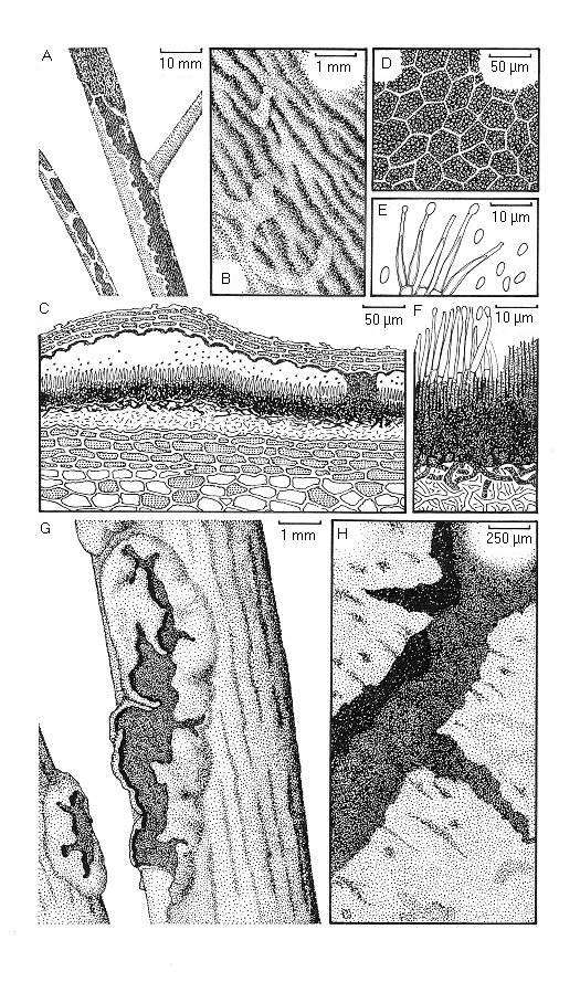
Cryptomyces maximus. A. Habit on twig. B. Conidiomatal surface viewed through a hand lens. C. Conidioma in vertical transverse section. D. Detail of conidiomatal upper wall in horizontal section, viewed with a compound microscope. E. Conidia and conidiogenous cells. F. Detail of lower part of conidioma in vertical transverse section. G. Ascomata viewed through a hand lens. H. Part of ascoma viewed with a dissecting microscope.