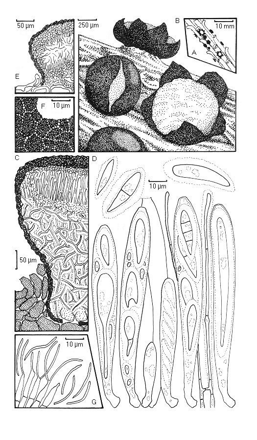
Tryblidiopsis pinastri. A. Habit on twig. B. Ascomata viewed with a dissecting microscope. C. Ascoma in vertical transverse section. D. Detail of ascomatal upper wall in horizontal section showing plate-like pattern. E. Asci, ascospores and paraphyses; most of the ascospores illustrated are atypical and probably the result of the in vitro maturation to which the illustrated specimen was subjected; the two free ascospores in the top left hand part of the illustration are typical of those observed in fresh naturally matured specimens. F. Conidioma in vertical transverse section. G. Conidiogenous cells and conidia.